What’s the Difference Between Head Scans?

Medical imaging can feel like alphabet soup — X-Ray, CT, MRI, MRA, PET. Each one has its own strengths, and together they give doctors a complete picture of what’s happening inside the head. Here’s the breakdown:
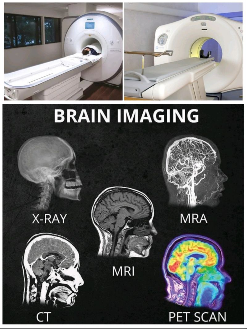
X-Ray – The quickest and most basic tool. Perfect for bones, it can show fractures or cracks in the skull. But it doesn’t reveal the brain itself.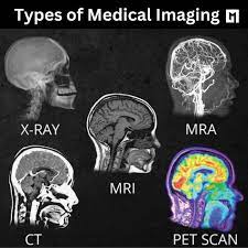
CT Scan – A rapid, powerful scan that works like an advanced X-ray. It captures images of the brain and is excellent in emergencies, spotting strokes, bleeds, or large tumors quickly. The tradeoff: less detail than MRI.
MRI – The gold standard for detail. It takes longer than a CT, but produces crystal-clear images of the brain’s structures. Doctors rely on it to detect small strokes, subtle bleeds, lesions, or early-stage tumors that a CT might miss.
MRA – A specialized type of MRI focused on blood vessels. It maps how blood flows through the brain, revealing blockages, narrowing, or aneurysms.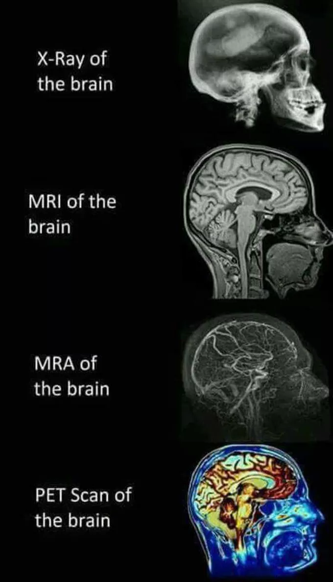
PET Scan – Unique because it shows function, not just structure. By tracking how much sugar different parts of the brain consume, PET scans highlight areas of high activity. Cancer cells, which burn energy rapidly, often glow the brightest — making PET a powerful tool for detecting and monitoring cancer.
In short:
X-Ray = Bone
CT = Quick brain snapshot
MRI = Detailed brain view
MRA = Blood flow
PET = Brain activity and cancer detection
Each scan reveals something the others can’t — and when combined, they give doctors the insight needed to save lives.